Damage from concussion alters the way information is transmitted between the two halves of the brain, according to a new study presented today at the annual meeting of the Radiological Society of North America (RSNA).
Research has shown that the corpus callosum, a bundle of nerve fibers that carries signals between the brain’s left and right hemispheres, is vulnerable to damage from mild traumatic brain injury, commonly known as concussion. Less is known about the impact of this damage on cognitive function.
To learn more, researchers at New York University (NYU) School of Medicine in New York City compared the condition of the corpus callosum in 36 patients with recent concussion to that of 27 healthy controls. They studied the participants’ brains with two innovative advances, including an MRI technique that uses measures of water diffusion to provide a microscopic view of the brain’s signal-carrying white matter.
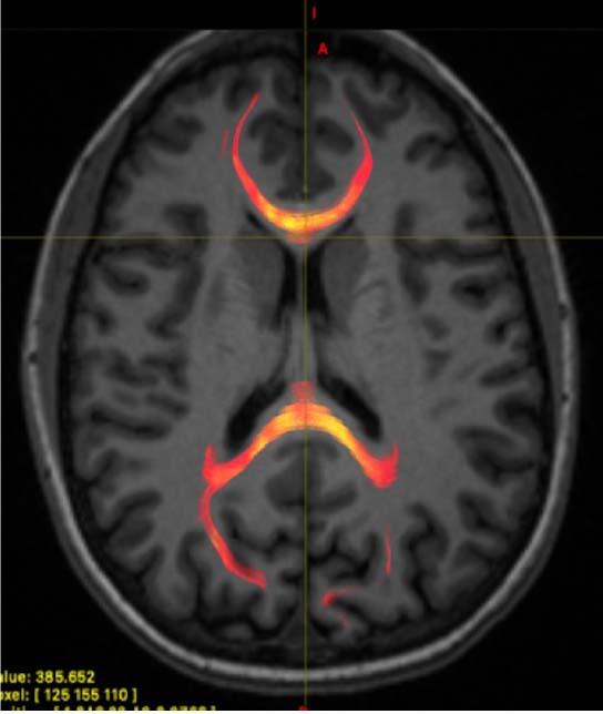
“Looking at how water molecules are diffusing in the nerve fibers in the corpus callosum and within the microenvironment around the nerve fibers allows us to better understand the white matter microstructural injury that occurs,” said study co-author Melanie Wegener, M.D., resident physician at NYU Langone Health in New York City.
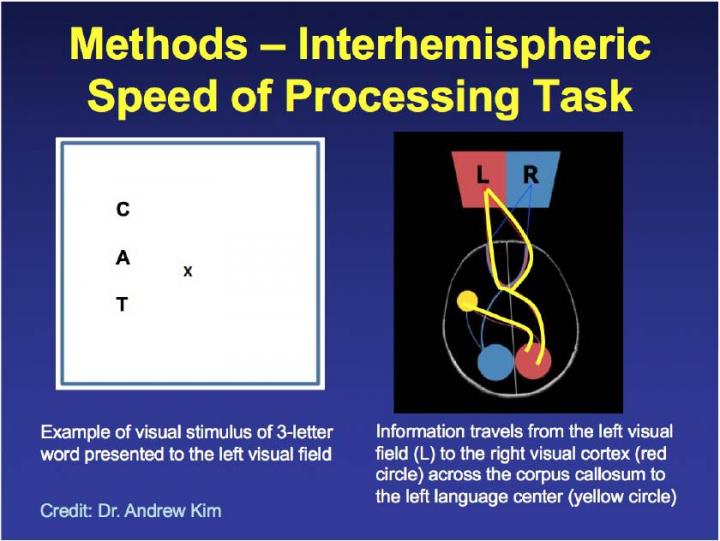
Dr. Wegener and colleagues combined the MRI findings with results from the study’s second innovative advance, called an Interhemispheric Speed of Processing Task, a test developed at NYU Langone that evaluates how well the two hemispheres in the brain communicate with each other.
[ad_336]
For the test, the participants were told to sit in a chair and focus their gaze on the letter X that was displayed on a screen directly in front of them. The researchers then flashed three-letter words to the right or the left of the X and asked the participants to say those words as quickly as possible. When the researchers evaluated this reaction time in both patients with concussion and healthy controls, they noticed an interesting phenomenon.
“There is a definite and reproducible delay in reaction time to the words presented to the left of the X compared with words presented to the right visual field,” Dr. Wegener said. “This shows it takes time for information to cross the corpus callosum from one hemisphere to the other, which is measured by the difference in response time between words presented to different sides of our visual field.”
This delay is likely due to the fact that language function is most often located in the brain’s left hemisphere. This means that information presented to the left visual field is first transmitted to the right visual cortex in the brain and then has to cross over the corpus callosum to get to the left language center. In contrast, words that are presented to the right visual field do not need to cross the corpus callosum.
[rand_post]
Performance on the test correlated with brain findings on MRI. In the healthy controls, reaction time corresponded with several diffusion measures in the splenium, an area of the corpus callosum located between the right visual cortex and the left language center. No such correlation was found in the concussion patients, suggesting microstructural changes relating to injury.
“We saw a correlation between white matter microstructure injury and the clinical status of the patient,” Dr. Wegener said. “This information could ultimately help with treatment in patients who have mild traumatic brain injury.”
For instance, Dr. Wegener said, patients could undergo MRI immediately after a concussion to see if they experienced any clinically important white matter injury and thus may benefit from early intervention.
“Another thing we can do is use MRI to look at patients’ brains during treatment and monitor the microstructure to see if there is a treatment-related response,” she said.